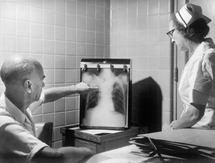
Pulmonary nodules are almost always incidental findings on a chest X-Ray that are either discovered accidentally or when looking for something else. With the advent of new screening programs for high risk adults with a previous smoking history, more than a million new cases of pulmonary nodules are found, with approximately a five percent malignancy rate. It is no wonder underwriters and physicians take these findings seriously, and a thorough work-up is done to be sure any nodule encountered has a benign outcome.
People with cough, suspected pneumonia, difficulty breathing, or an abnormal lung field exam on physical are obvious candidates for X-Rays that may discover a lung nodule. In addition, the US Preventative Services Task Force (USPSTF) now recommends annual screening for lung cancer with low dose CT scanning in adults aged 50-80 who have a 20 pack year history and are either current smokers or who have quit smoking in the previous 15 years. The objective evidence is striking for this testing: findings showed a 20 percent reduction in lung cancer related mortality as a result of the scanning.
While multiple nodules may involve a more systemic process such as fungal infection, sarcoid, bacterial infection or tuberculosis, the single pulmonary nodule is most concerning as the risk of malignancy is significant. The growth may be a primary cancer or a secondary malignancy (metastasis) from a different body organ, and work-ups to try to determine the etiology as the only certain method of distinguishing a malignant from a benign process is by biopsy, which is often invasive.
The risk of malignancy is highest in solid nodules that are large sized, have calcifications that are not symmetric, and that double in size between one month and one year of observation. Nodules that grow more quickly are more likely inflammatory or infectious, and a different cause should be sought. Other characteristics of suspicious nodules include irregular or spiculated borders, ground glass appearance on X-Ray, and location in the upper lung lobes. Increasing age and cigarette smoking are associated with higher risk of lung cancer.
Work-up of the nodule is done in a sequential manner. If discovered on X-Ray, a CT scan is usually the next step to identify exact size and characteristics. Smaller lesions are generally monitored with serial CT scans. If a generalized process is suspected, that is worked up at the same time. Bronchoscopy can help make a diagnosis with direct visualization and cell washings from the suspicious area. CT guided biopsies are often sufficient for a definitive diagnosis, but open lung biopsies may have to be obtained when the lesion is small or in a spot that is inaccessible to the bronchoscope or to getting tissue in a vulnerable area.
The recommended management of an incidentally detected solid pulmonary nodule as defined by CHEST (the American College of Chest Physicians) recommends follow-up based on nodule size and intermediate and high risk factors that combine the aforementioned characteristics favoring malignancy. Nodule biopsies and bronchoscopy are considered when the nodule is within the reach of either procedure. When the risk of malignancy is significantly high, surgical resection is the procedure of choice. Nonsurgical options like ablative therapy or stereotactic radiotherapy may be considered for those who are at high risk of complication or death from a resection.
Underwriting a case with a pulmonary nodule starts with the results of investigation—a new nodule has to be worked up and evaluated before any case can proceed. There should be a good description of the nodule or nodules, including size, consistency, shape and margins from the original study. There should be follow-up of the nodule to see if it has been increasing in size, and how long that interval is. Biopsy and bronchoscopy results and the work-up from the chest physician and/or surgeon should have detailed and inclusive notes. If the nodule is for a more generalized cause, that should also have been worked up and treated.
A solitary pulmonary nodule is never a welcome and most often an unanticipated finding, but the good news is that most turn out to be benign lesions or part of a more generalized treatable cause. Either way, results from a detailed investigation are necessary for a good and expedient case outcome.


